What is the Cornea?
The cornea is your eye’s outermost layer. It is the clear, dome-shaped surface that covers the front of the eye. To see well, the cornea must remain transparent to refract light properly and all layers of the cornea must be free of any cloudy or opaque areas. When there is a problem with your cornea, it can significantly affect your vision.
Understanding the Structure of Your Cornea
Although the cornea is clear and seems to lack substance, it is actually a highly organized group of cells and proteins. Unlike most tissues in the body, the cornea contains no blood vessels to nourish or protect it against infection. Instead, the cornea receives its nourishment from the tears and aqueous humor (a fluid in the anterior portion of the eye) that fills the chamber behind it. The cornea must remain transparent to refract light properly, and the presence of even the tiniest blood vessels can interfere with this process.
The corneal tissue is arranged in five basic layers, each having an important function. These five layers are:
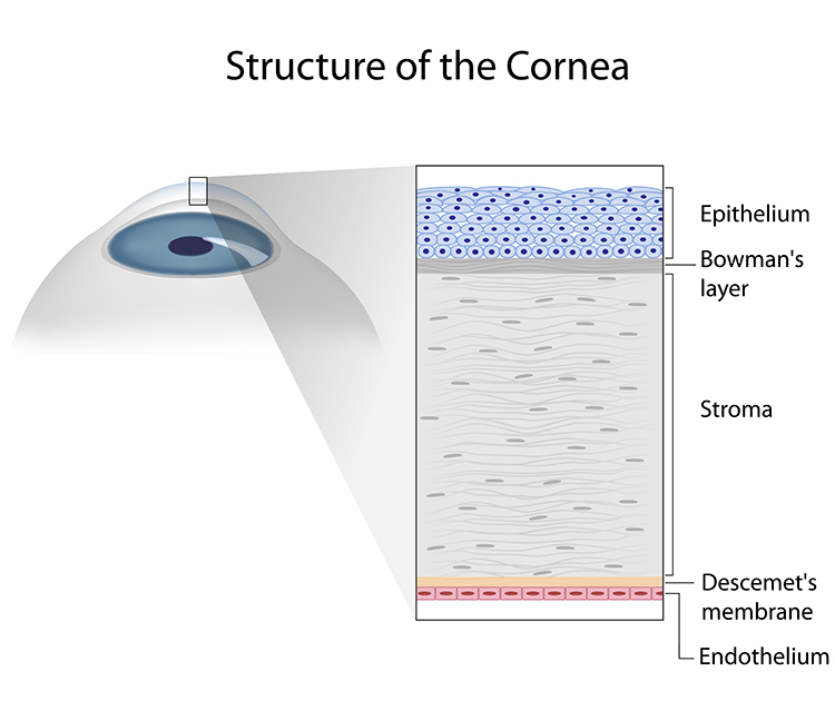
The Epithelium
The epithelium is the cornea’s outermost region, comprising about 10 percent of the tissue’s thickness. The epithelium functions primarily to: (1) block the passage of foreign material, such as dust, water, and bacteria, into the eye and other layers of the cornea; and (2) provide a smooth surface that absorbs oxygen and cell nutrients from tears, then distributes these nutrients to the rest of the cornea. The epithelium is filled with thousands of tiny nerve endings that make the cornea extremely sensitive to pain when rubbed or scratched. The part of the epithelium that serves as the foundation on which the epithelial cells anchor and organize themselves is called the basement membrane.
Bowman’s Layer
Lying directly below the basement membrane of the epithelium is a transparent sheet of tissue known as Bowman’s layer. It is composed of strong layered protein fibers called collagen. Once injured, Bowman’s layer can form a scar as it heals. If these scars are large and centrally located, some vision loss can occur.
Stroma
Beneath Bowman’s layer is the stroma, which comprises about 90 percent of the cornea’s thickness. It consists primarily of water (78 percent) and collagen (16 percent), and does not contain any blood vessels. Collagen gives the cornea its strength, elasticity, and form. The collagen’s unique shape, arrangement, and spacing are essential in producing the cornea’s light-conducting transparency.
Descemet’s Membrane
Under the stroma is Descemet’s membrane, a thin but strong sheet of tissue that serves as a protective barrier against infection and injuries. Descemet’s membrane is composed of collagen fibers (different from those of the stroma) and is made by the endothelial cells that lie below it. Descemet’s membrane is regenerated readily after injury.
Endothelium
The endothelium is the extremely thin, innermost layer of the cornea. Endothelial cells are essential in keeping the cornea clear. Normally, fluid leaks slowly from inside the eye into the middle corneal layer (stroma). The endothelium’s primary task is to pump this excess fluid out of the stroma. Without this pumping action, the stroma would swell with water, become hazy, and ultimately opaque. In a healthy eye, a perfect balance is maintained between the fluid moving into the cornea and fluid being pumped out of the cornea. Once endothelium cells are destroyed by disease or trauma, they are lost forever. If too many endothelial cells are destroyed, corneal edema and blindness ensue, with corneal transplantation the only available therapy.
If you have been diagnosed with a corneal problem, schedule an appointment with Wilmington Eye and our corneal specialist.
The Function of Your Cornea
The cornea serves two main functions:
- It helps to shield the rest of the eye from germs, dust, and other harmful matter. The cornea shares this protective task with the eyelids, the eye socket, tears, and the white part of the eye (sclera).
- The cornea acts as the eye’s outermost lens. It functions like a window that controls and focuses the entry of light into the eye. The cornea contributes between 65-75 percent of the eye’s total focusing power.
When light strikes the cornea, it bends–or refracts–the incoming light onto the lens. The lens further refocuses that light onto the retina, a layer of light-sensing cells lining the back of the eye that starts the translation of light into vision. For you to see clearly, light rays must be focused by the cornea and lens to fall precisely on the retina. The retina converts the light rays into impulses that are sent through the optic nerve to the brain, which interprets them as images.
The refractive process is similar to the way a camera takes a picture. The cornea and lens in the eye act as the camera lens. The retina is similar to the film or digital sensor. If the image is not focused properly, the film (or retina) receives a blurry image.
If you are experiencing an issue with your vision, schedule an appointment with our doctors today.